Bones In Leg Diagram / 11. Orthopedic Surgery | Basicmedical Key : 12 photos of the bones leg diagram picture.
byAdmin•
0
Bones In Leg Diagram / 11. Orthopedic Surgery | Basicmedical Key : 12 photos of the bones leg diagram picture.. Femur bone structure royalty free vector image. Learn vocabulary, terms and more with flashcards, games and other study tools. When your muscles contract, they pull the bone they're. Your leg bones are very large and strong to help support the weight of your body. Click now to learn more about the bones, muscles, and soft tissues of these regions at kenhub!
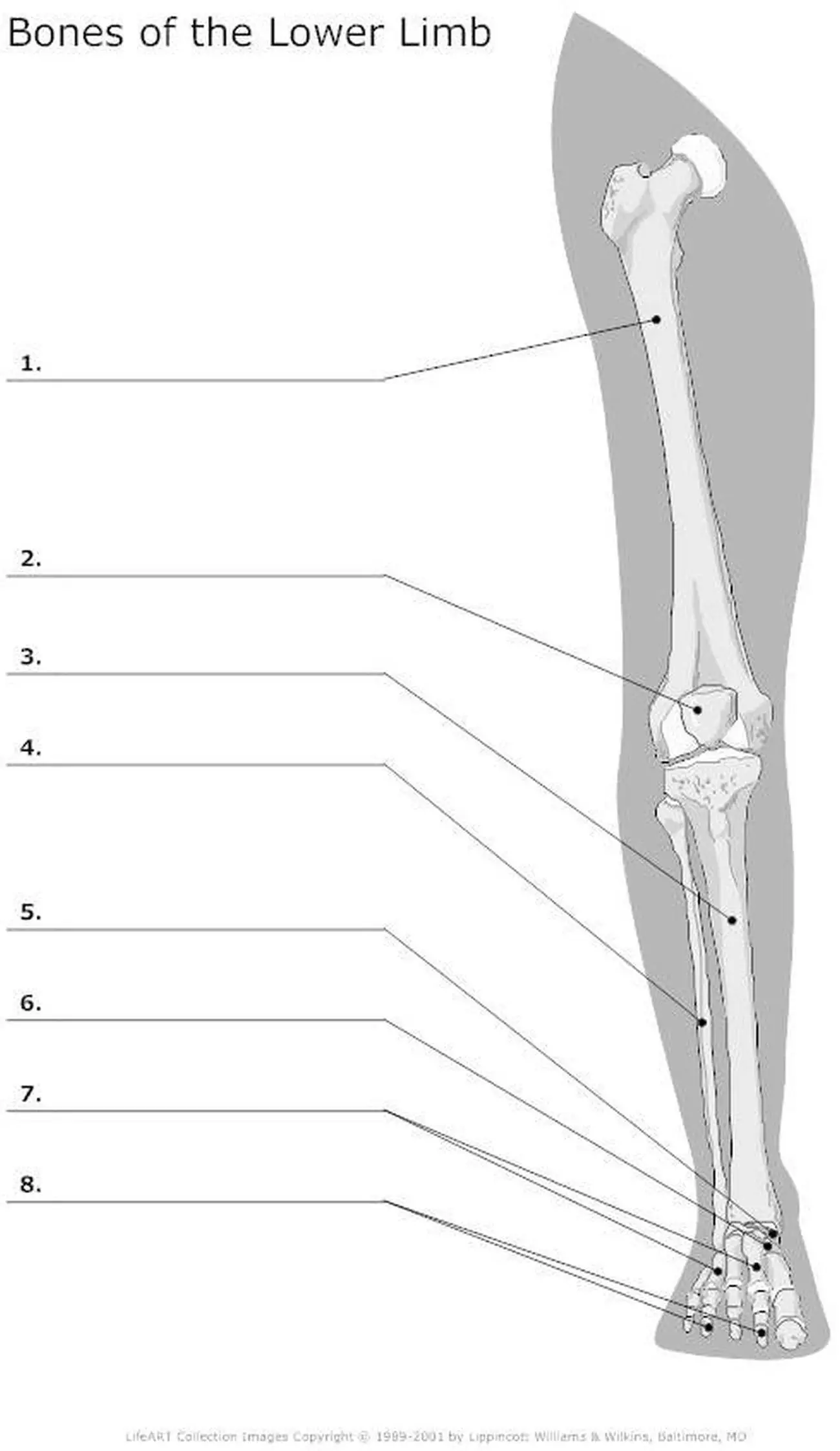
Diagram of bones 16 best bones in the leg images on pinterest antique 1890s medical anatomy diagram leg bones skeleton He leg's main function in the human is for locomotion and support of the rest of the body. Posted on january 20, 2015 by admin. This diagram depicts diagram leg bones anatomy. The foot bones shown in this diagram are the talus, navicular, cuneiform, cuboid, metatarsals and calcaneus.
leg muscles diagram - Free Large Images from www.free-largeimages.com Master leg and knee anatomy using our topic page. The bones of your leg have roughened patches on their surfaces where muscles are attached. The bone that goes from your pelvis to your knee is called the femur (say: License image the bones of the leg are the femur, tibia, fibula and patella. Want to learn more about it? When your muscles contract, they pull the bone they're. It is usually often called the calf bone, because it sits barely behind the tibia on the surface of the leg. The foot bones shown in this diagram are the talus, navicular, cuneiform, cuboid, metatarsals and calcaneus.
The wires will probably be coloured similar to the actual wires you can be employing.
The wires will probably be coloured similar to the actual wires you can be employing. Top suggestions for human leg bones diagram. Nervsystemet anatomy, diagram & function | health. Click and start learning now! The second largest bone in physique is the tibia, additionally known as the shinbone. Bones pain hand and arm bones diagram. The foot bones shown in this diagram are the talus, navicular, cuneiform, cuboid, metatarsals and calcaneus. When your muscles contract, they pull the bone they're. Electrical wiring diagrams diagram of lower limb bones that happen to be in shade have an advantage more than ones that happen to be black and white only. 12 photos of the bones leg diagram picture. He leg's main function in the human is for locomotion and support of the rest of the body. Your leg bones are the longest and strongest bones in your body. Learn vocabulary, terms and more with flashcards, games and other study tools.
Want to learn more about it? 12 photos of the bones leg diagram picture. Nasal bone anatomy x ray 12 photos of the nasal bone anatomy x ray nasal bone. He leg's main function in the human is for locomotion and support of the rest of the body. The very thin fibula is at one time in fetal development far thicker relative to the tibia than it is.
Osteology Unit - M.Y. Online Portfolio from mkyousif17.weebly.com A) that they shared a common ancestor. At the distal end of the femur, two rounded condyles meet the tibia and fibula bones of the lower leg to form the knee joint. The bones of the leg are the femur, tibia, fibula and patella. Human anatomy diagrams show internal organs, cells, systems, conditions, symptoms and sickness information and/or tips for healthy living. What does this suggest about mammals? The human leg consists of 8 bones, 4 per leg. The bones involved in it, however, are only the femur and the tibia, although the smaller bone of the leg, the fibula, is carried along in the movements of flexion, extension, and slight rotation that this joint permits. The bones of the leg are the femur, tibia, fibula and patella.
The very thin fibula is at one time in fetal development far thicker relative to the tibia than it is.
The femur, or thigh bone, is the largest, heaviest, and strongest bone in the human body. Related posts of diagram of leg bones nasal bone anatomy x ray. The bone that goes from your pelvis to your knee is called the femur (say: Click and start learning now! The foot bones shown in this diagram are the talus, navicular, cuneiform, cuboid, metatarsals and calcaneus. Human anatomy diagrams show internal organs, cells, systems, conditions, symptoms and sickness information and/or tips for healthy living. He leg's main function in the human is for locomotion and support of the rest of the body. Top suggestions for human leg bones diagram. The bones of your leg have roughened patches on their surfaces where muscles are attached. This diagram depicts diagram leg bones anatomy. 12 photos of the bones leg diagram picture. What does this suggest about mammals? An electrical wiring diagram can be as simple as a diagram demonstrating how to set up a fresh swap with your hallway.
When you stand or walk, all the weight of your upper body rests on them. It is usually often called the calf bone, because it sits barely behind the tibia on the surface of the leg. Explore more like human leg bones diagram. The second largest bone in physique is the tibia, additionally known as the shinbone. Master leg and knee anatomy using our topic page.
Pictures Of Bones Of The Lower Extremities from healthiack.com Bones pain hand and arm bones diagram. The bone that goes from your pelvis to your knee is called the femur (say: Top suggestions for human leg bones diagram. Learn vocabulary, terms and more with flashcards, games and other study tools. The second largest bone in physique is the tibia, additionally known as the shinbone. 12 photos of the bones leg diagram picture. An electrical wiring diagram can be as simple as a diagram demonstrating how to set up a fresh swap with your hallway. Click and start learning now!
The human leg consists of 8 bones, 4 per leg.
The foot bones shown in this diagram are the talus, navicular, cuneiform, cuboid, metatarsals and calcaneus. The knee is a strong but flexible hinge joint. Electrical wiring diagrams diagram of lower limb bones that happen to be in shade have an advantage more than ones that happen to be black and white only. Editor · aug 13, 2017 ·. The bones of the leg are the femur, tibia, fibula and patella. Posted on january 20, 2015 by admin. Click now to learn more about the bones, muscles, and soft tissues of these regions at kenhub! Related posts of diagram of leg bones nasal bone anatomy x ray. The bones of the leg are the femur, tibia, fibula and patella. They are extensively utilized in industries. 12 photos of the bones leg diagram picture. The bone that goes from your pelvis to your knee is called the femur (say: When you stand or walk, all the weight of your upper body rests on them.